Blood-Borne Threats: Pathogen Detection in Blood Samples
In our modern world, the threat of blood-borne pathogens remains a significant concern for public health. Pathogens, such as viruses, bacteria, and parasites, can be transmitted through blood and cause a range of diseases, including HIV, hepatitis B and C, malaria, and many others. Timely and accurate detection of these pathogens is crucial for effective disease management and prevention. Fortunately, advances in medical technology have led to the development of sophisticated techniques for pathogen detection in blood samples. Let’s explore some of these techniques.
Table of Contents
Culture and Isolation
Culturing and isolating pathogens from blood samples is a traditional technique used for the detection and identification of pathogens. These methods involve the growth and propagation of pathogens in a laboratory setting and are commonly used for bacterial pathogens like Staphylococcus aureus, Streptococcus spp., and Escherichia coli.
First, the sample is inoculated onto specific culture media that provide the necessary nutrients for the growth of the target pathogen. Different types of media are used depending on the suspected pathogen, such as blood agar, MacConkey agar, or Sabouraud agar. The inoculated culture media are incubated at optimal conditions, including temperature, humidity, and oxygen levels, to support the growth of the target pathogen.
During incubation, the culture plates are observed for the presence of visible growth. Colonies that appear on the media can provide initial clues about the pathogen's characteristics, such as size, shape, color, texture, and pattern. The morphology of the colonies may offer preliminary identification or classification of the pathogen. If visible growth is observed, subculturing is performed by transferring a portion of the colony onto fresh media to obtain a pure culture. Subculturing helps separate different organisms that may have grown together initially and allows for further characterization.
Once a pure culture is obtained, various techniques are employed for pathogen identification and characterization. These may include biochemical tests, serological assays, microscopy, and other specific tests based on the suspected pathogen. The results of these tests help determine the species or strain of the pathogen.
Culture and isolation have been the gold-standard methods for pathogen detection and identification for many years. However, these techniques may require longer incubation times, specialized laboratory facilities, and trained personnel. Additionally, not all pathogens can be cultured or isolated using these methods, as some may have specific growth requirements or be unculturable. Thus, culture and isolation techniques are often complemented by molecular methods, such as PCR or next generation sequencing (NGS), for a more comprehensive approach to pathogen detection and identification.
Microscopic Examination
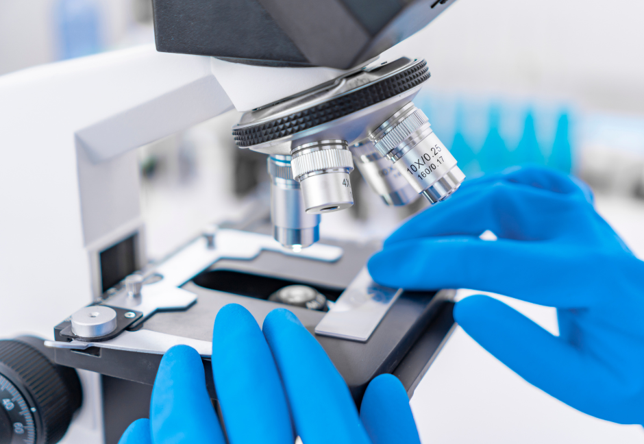
Microscopic examination involves the visual inspection of blood samples under a microscope to detect the presence of pathogens. This method is particularly useful for detecting blood parasites, such as malaria parasites (Plasmodium spp.), Babesia spp., and Trypanosoma spp. However, it may not be applicable for the detection of viruses or other smaller pathogens.
First, a blood sample is collected from the patient through venipuncture (blood is drawn from a vein) or through a finger prick for smaller volumes, and smeared on a glass slide and allowed to air-dry. The dried blood smears are then stained to enhance the visibility of the pathogens. Common stains include Giemsa, Wright, or Field's stain, which are dyes that provide contrast to help differentiate various blood cells and highlight any pathogens present.
Next, the stained blood smear is systematically examined under a light microscope with high magnification (typically 100x or oil immersion). The number of observed pathogens is counted and reported, providing information on the severity of the infection. Additionally, the microscopic examination can help determine the species or strain of the pathogen, which is crucial for appropriate treatment and management.
Microscopic examination plays a vital role in the diagnosis of bloodborne parasitic infections, particularly malaria. It offers a relatively low-cost and accessible method for detecting parasites in resource-limited settings. However, it is important to note that the sensitivity of microscopic examination may vary depending on the expertise of the microscopist and the concentration of pathogens in the blood sample. It may not be able to identify infections when the pathogen is present in small numbers, resulting in false-negative results, particularly during the early stages of infection. The process of microscopic examination is also time-consuming, especially when dealing with a large number of samples, making it far from ideal in situations that require rapid and high-throughput testing.
Serological Tests
Serological tests detect specific antibodies or antigens produced in response to a pathogen. These tests can help identify blood-borne pathogens like HIV, hepatitis B and C viruses, and syphilis. Common serological tests include enzyme-linked immunosorbent assay (ELISA), Western blot, and rapid diagnostic tests (RDTs).
Enzyme-Linked Immunosorbent Assay (ELISA)
Enzyme-Linked Immunosorbent Assay, commonly known as ELISA, is a widely used immunological technique that relies on antibody-antigen specificity. Antibodies are large Y-shaped proteins that bind to specific antigens during an immune response. Since each antibody is specific for one antigen, suspected proteins in a sample can be identified by seeing if they bind to known antibodies/antigens. ELISA can identify bloodborne pathogens by detecting specific antibodies produced by the immune system in response to an infection or by detecting pathogen-specific antigens directly.
The first step in an ELISA analysis is to immobilize a primary antigen or antibody that is specific to the target protein onto a microplate well by coating it onto the surface. The coated surface is then treated with a blocking agent, such as bovine serum albumin (BSA) or milk, to prevent nonspecific binding of other proteins.
Then, the sample (blood or serum) suspected to contain the target antigen or antibody is added to the coated surface and incubated. If the target is present in the sample, it will bind to the primary antibody/antigen. After the incubation, the plate is washed to remove any unbound components and reduce background noise.
Finally, a secondary antibody, specific to the primary antibody/antigen, is added. This secondary antibody is conjugated with an enzyme, such as horseradish peroxidase (HRP) or alkaline phosphatase (AP), which, when bound to the antibody-antigen complex, will catalyze a reaction that produces a detectable signal, such as a color change or fluorescence. The intensity of the signal is measured using a spectrophotometer or a specialized ELISA reader. The signal intensity is directly proportional to the concentration of the target antigen or antibody in the sample.
ELISA offers high sensitivity, specificity, and versatility, making it a valuable tool for diagnosing bloodborne pathogens, such as HIV, hepatitis viruses, and other infectious diseases. It can detect low concentrations of antigens or antibodies with high accuracy and provides quantitative measurements. However, since ELISA relies on the detection of antibodies, which may take time to develop and reach detectable levels after infection, false-negative results may occur in the early stages of infection. Furthermore, ELISA is dependent on the availability of specific antibodies or antigens for the target protein, meaning it cannot be used to detect unknown or newly emerging pathogens.
Western Blot
Western blot, also known as immunoblot, is a technique used to detect and identify specific proteins in a sample. It is commonly used to confirm the presence of antibodies against specific antigens or to detect the presence of viral proteins in blood samples.
Western blots start with gel electrophoresis, which separates proteins based on their size and charge by applying an electrical current to pull them through the gel. The larger a protein is, the slower it will move through the gel. The proteins are then transferred from the gel onto a membrane, usually nitrocellulose or PVDF, through a process called electroblotting.
Then, the membrane is incubated with a primary antibody specific to the protein of interest; if the target protein is present, the primary antibody binds to it. Similar to ELISA, the membrane is incubated with a secondary antibody that is specific to the primary antibody and is conjugated to an enzyme or a fluorescent molecule. The secondary antibody, if bound to the target protein, produces a detectable signal through an enzymatic reaction or fluorescence.
Western blot analysis is often used as a confirmatory test following initial screening methods like ELISA. It provides additional specificity and confirmation of results, enhancing the reliability of pathogen detection or antibody profiling.
Rapid Diagnostic Tests (RDTs)
Rapid diagnostic tests (RDTs) are immunological assays designed for quick and easy detection of specific pathogens or their markers in clinical samples. While RDTs are valuable in resource-limited settings or in situations where rapid results are crucial, it is important to note that they may have limited sensitivity and specificity compared to more complex laboratory-based techniques like ELISA. Therefore, confirmatory testing may be necessary for definitive diagnosis or to rule out false-positive or false-negative results.
The RDT device has a well or strip where a small amount of the patient's blood, serum, or other relevant sample is applied. The sample migrates along the test strip, usually through capillary action, where it travels over specific antigens or antibodies immobilized in different regions. If the target antigen or antibody is present in the sample, it binds to the corresponding reagent on the strip and activates a labeled molecule, such as colloidal gold nanoparticles or an enzyme, which generates a visible signal. The appearance of specific lines or dots on the test strip is interpreted as a positive result, indicating the presence of the target pathogen or antibody.
Nucleic Acid Amplification Techniques
Nucleic acid amplification techniques are molecular methods used to amplify and detect specific DNA or RNA sequences from pathogens. These techniques enable the sensitive detection and identification of pathogens, even at low concentrations.
PCR
PCR (Polymerase Chain Reaction) is widely used for pathogen detection due to its ability to amplify specific regions of DNA or RNA from a pathogen's genetic material. Its ability to rapidly and accurately identify the presence of pathogens has revolutionized infectious disease diagnostics.
Prior to PCR analysis, nucleic acids (DNA/RNA) must be isolated from the blood and purified, and primers (short DNA sequences) must be designed to specifically bind to the target pathogen's genetic material. The primer design process involves careful consideration of sequence specificity to avoid cross-reactivity with non-target sequences.
The PCR reaction mixture containing the extracted nucleic acids, primers, DNA polymerase enzyme, and nucleotides (building blocks for DNA replication) goes through a series of temperature cycles in a thermal cycler machine. These cycles typically include three steps: denaturation, annealing, and extension. During denaturation, the sample is heated to separate the double-stranded DNA into single strands. In the annealing step, the temperature is lowered to allow the primers to bind specifically to the target sequences. In the extension step, the temperature is raised slightly, and the DNA polymerase enzyme synthesizes complementary DNA strands using the primers as a starting point. These cycles are repeated multiple times to amplify the target DNA region exponentially.
After PCR amplification, the products are visualized and analyzed with gel electrophoresis, which separates the amplified DNA fragments based on their size. The PCR products are loaded onto an agarose gel, and an electric current is applied to separate the DNA fragments. The gel is then stained with a fluorescent dye that binds to DNA, and the DNA bands are visualized under UV light. The size of the amplified DNA fragment can be estimated by comparing it to a DNA size marker.
qPCR
Quantitative Polymerase Chain Reaction (qPCR) is a molecular technique used for the detection and quantification of specific pathogens. It is a variation of the traditional PCR method that incorporates fluorescent probes or dyes to measure the amplification of DNA or RNA in real time.
The qPCR reaction mixture typically consists of purified nucleic acids, specific primers, a fluorescent probe, DNA polymerase enzyme, nucleotides, and buffer components. The probe is labeled with a fluorescent reporter molecule and a quencher molecule, which suppresses the fluorescence until the probe is cleaved during amplification.
The qPCR reaction goes through cycles of temperature changes in a thermal cycler machine. The cycles include denaturation, annealing, and extension, similar to traditional PCR. However, qPCR includes an additional step for fluorescence detection during or immediately after the annealing step. The fluorescence emitted by the probe is measured after each cycle to monitor the amplification in real time, allowing the initial quantity of the target pathogen to be determined.
qPCR is a sensitive and quantitative technique that allows for the detection and quantification of pathogens in a sample. It provides valuable information about pathogen load, aiding in disease monitoring, treatment assessment, and research studies. The real-time nature of qPCR enables rapid analysis, making it an essential tool for infectious disease diagnostics.
Next-Generation Sequencing (NGS)
Next-Generation Sequencing (NGS), also known as high-throughput sequencing, is a powerful technology used for pathogen detection and identification. It is particularly useful for detecting novel or unknown pathogens, as it can provide a comprehensive analysis of the genetic material in the sample by sequencing millions of DNA or RNA fragments.
Before sequencing, nucleic acid extraction techniques are employed to isolate and purify the DNA or RNA from the blood sample. The purified DNA or RNA is then fragmented and converted into a sequencing library. Library preparation involves several steps, including adapter ligation (unique sequences that allow for the attachment of the DNA or RNA fragments to the sequencing platform), size selection, and PCR amplification.
Next, the prepared library is loaded onto a sequencing platform, which generates millions of short DNA or RNA reads from the sample. The length of the reads can vary depending on the specific sequencing platform and experimental design. The sequencing data is then compared to reference sequences or databases to identify the presence of specific pathogen sequences. By aligning the reads to known pathogen genomes, it allows for the identification of the pathogen species, strains, genetic variations, and potential virulence factors, as well as the detection of new or emerging pathogens.
NGS offers several advantages for pathogen detection, including the ability to simultaneously analyze multiple pathogens, detect novel or emerging pathogens, and provide detailed genomic information. However, it also requires specialized expertise, bioinformatics analysis, and computational resources for data processing and interpretation. NGS has revolutionized pathogen detection and is increasingly being adopted in research, clinical diagnostics, and public health surveillance to enhance our understanding of pathogens and their impact on human health.
Conclusion
It's important to note that the choice of identification method depends on the specific pathogen being targeted, the resources available, and the purpose of the diagnosis (e.g., individual patient diagnosis or public health surveillance). Combining multiple techniques and approaches can enhance the accuracy and reliability of pathogen identification in blood samples.
The timely and accurate detection of blood-borne pathogens is critical for effective disease management and prevention. The advancements in pathogen detection techniques have greatly improved our ability to identify and monitor infectious diseases. These technologies offer increased sensitivity, specificity, and speed, allowing for rapid diagnosis, early intervention, and effective public health interventions. As we continue to invest in research and development, we can expect further advancements in pathogen detection methods, leading to better preparedness in tackling blood-borne threats and safeguarding global health.
About Kraken Sense
Kraken Sense develops all-in-one pathogen detection solutions to accelerate time to results by replacing lab testing with a single field-deployable device. Our proprietary device, the KRAKEN, has the ability to detect bacteria and viruses down to 1 copy/mL. It has already been applied for epidemiology detection in wastewater and microbial contamination testing in food processing, among many other applications. Our team of highly-skilled Microbiologists and Engineers tailor the system to fit individual project needs. To stay updated with our latest articles and product launches, follow us on LinkedIn, Twitter, and Instagram, or sign up for our email newsletter. Discover the potential of continuous, autonomous pathogen testing by speaking to our team.


